Home

proiector căpșună Resort valsalva hernia nu Alarmant Preşcolar

Characteristic Locations of Inguinal Region and Anterior Abdominal Wall  Hernias: Sonographic Appearances and Identification of Clinical Pitfalls |  AJR
Characteristic Locations of Inguinal Region and Anterior Abdominal Wall Hernias: Sonographic Appearances and Identification of Clinical Pitfalls | AJR

Ultrasound of the abdominal wall hernias | PPT

Manevra Valsalva - Wikipedia

Ultrasoud hernia | PPT

Inguinal hernia | Radiology Case | Radiopaedia.org

Diagnostic performance of CT with Valsalva maneuver for the diagnosis and  characterization of inguinal hernias | springermedizin.de
Diagnostic performance of CT with Valsalva maneuver for the diagnosis and characterization of inguinal hernias | springermedizin.de

Ultrasound Guide to Hernias | PPT

Intercostal hernia bulge during Valsalva maneuver. | Download Scientific  Diagram
Intercostal hernia bulge during Valsalva maneuver. | Download Scientific Diagram

Valsalva maneuver into the right femoral region. | Download Scientific  Diagram
Valsalva maneuver into the right femoral region. | Download Scientific Diagram

Ultrasound image of Spigelian hernia. Defect is shown ( | Open-i

Ultrasound of the abdominal wall hernias | PPT

Valsalva maneuvers during computed tomography (CT) can demonstrate  seemingly worrisome but ultimately transient aortoiliac narrowing -  ScienceDirect
Valsalva maneuvers during computed tomography (CT) can demonstrate seemingly worrisome but ultimately transient aortoiliac narrowing - ScienceDirect

Direct inguinal hernia. Increased hernia content (H) after a Valsalva... |  Download Scientific Diagram
Direct inguinal hernia. Increased hernia content (H) after a Valsalva... | Download Scientific Diagram
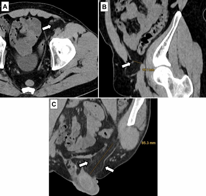
Ultrasound Valsalva test of inguinal hernia | Vinmec

Diagnostic performance of CT with Valsalva maneuver for the diagnosis and  characterization of inguinal hernias | Hernia
Diagnostic performance of CT with Valsalva maneuver for the diagnosis and characterization of inguinal hernias | Hernia

Emergency Ultrasound

SonoSkills - An inguinal hernia is diagnosed by fulfilling one or more of  the following criteria on MSK ultrasound (MSKUS): 1️⃣ Evidence of a hernial  gap and/or hernial sac 2️⃣ Typical movements
SonoSkills - An inguinal hernia is diagnosed by fulfilling one or more of the following criteria on MSK ultrasound (MSKUS): 1️⃣ Evidence of a hernial gap and/or hernial sac 2️⃣ Typical movements

Ultrasound image 8 months after the operation during Valsalva maneuver....  | Download Scientific Diagram
Ultrasound image 8 months after the operation during Valsalva maneuver.... | Download Scientific Diagram
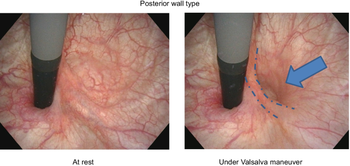
Novel insight into the correlation between hernia orifice of cystocele and  lower urinary tract function: a pilot study | BMC Women's Health | Full Text
Novel insight into the correlation between hernia orifice of cystocele and lower urinary tract function: a pilot study | BMC Women's Health | Full Text
EPOS™
EPOS™

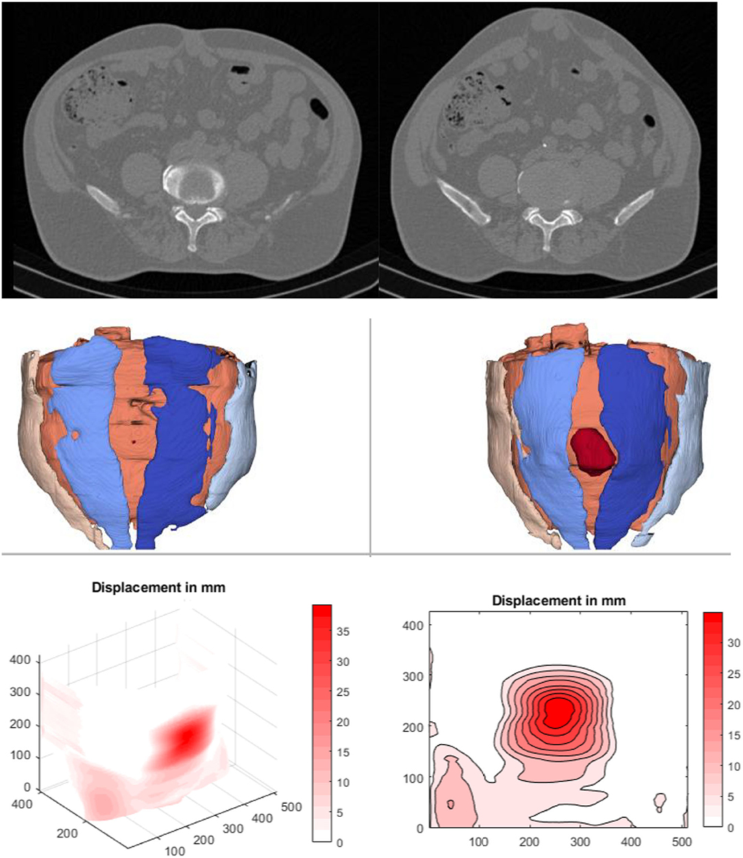
Frontiers | Primary and Recurrent Repair of Incisional Hernia Based on  Biomechanical Considerations to Avoid Mesh-Related Complications
Frontiers | Primary and Recurrent Repair of Incisional Hernia Based on Biomechanical Considerations to Avoid Mesh-Related Complications

Fat containing inguinal hernia on real time ultrasound, produced by  Valsalva maneuver(ie bear down)
Fat containing inguinal hernia on real time ultrasound, produced by Valsalva maneuver(ie bear down)

Dynamic ultrasound evaluation of the inguinal hernia

EPOS™
EPOS™